Please note: this page contains images of human skeletal remains.
Overview:
Since 1996, the Sedgeford Historical and Archaeological Research Project (SHARP) has excavated 291 whole or partial articulated skeletons from Boneyard, an Anglo-Saxon cemetery in Sedgeford, along with significant quantities of disarticulated human bone. A further 126 skeletons from the same cemetery, excavated in the 1950s and 60s, have also been identified in other collections. Post-excavation analysis is currently being carried out on the entire extant population (Faulkner et al 2014:138). From the remains of the ancient Sedgeford people we have learned a great deal about past lives and experiences. The Introduction to Human Remains course delivered by SHARP gave me an exceptional opportunity to learn how to carry out the archaeological interpretation of ancient human remains and prepare an osteological report.
The course covered basic anatomy, determination of age and sex, dentition and paleopathology, as well as recording methods. The ethical aspect of studying human remains and how to interpret population data was also discussed. This allowed me the opportunity to study human skeletal remains using skeletons and disarticulated bones excavated from the Boneyard site. The program was delivered by five experts in the field of osteology over a packed six-day period and we were examined on three occasions on what we had learned. Each test had us identifying various human bones in various states of preservation. We were asked to identify the bone, its orientation, the sex, age, relevant features, and any pathology or non-metric traits. The week was based around the recording of an individual skeletal assemblage, in my case that of skeleton number S1016. To achieve this the week was broken down into a series of lectures, seminars, and practical sessions which mirrored the relevant sections within the osteological report.
According to Simon Mays (Mays 2010:46) the purpose of an osteological report is to:
- Shed light on research questions pertinent to the site and the region in which it is situated
- Make osteological data available to the wider scientific community
- Alert other researchers to the existence of the material
- Act as a guide for researchers wishing to study the material.
The recording for each skeletal assembly, and that of disarticulated bone, at SHARP is part of a wider research project into the Anglo-Saxon inhabitants of Sedgeford. The report discussed here focuses on one skeleton which was identified as ‘of interest’ due to the osteological indications that the individual had suffered a great deal of head trauma around the time of death. The following is a breakdown of the osteological recording for my assigned skeleton completed during the course followed by an interpretation of the findings relating to the individual’s death. It is the cause of death in this instance which is the focus of the research question.
Sedgeford Historical and Archaeological Research Project (SHARP)
SHARP is located in the small village of Sedgeford which is in the north west of Norfolk, England. It has been an active research project since 1996 and the excavations conducted there span the Late Mesolithic, Late Neolithic/Early Bronze Age, Iron Age, Romano-British, Anglo-Saxon, Medieval, and WW1 periods. The project runs a series of courses over a six week period during the summer. These cover environmental archaeology, archaeometallurgy, non-invasive archaeology, and human remains. More information on the research conducted by the project and the courses offered can be found on their website here. Excavation volunteers and course participants tend to camp onsite and there are numerous facilities available to make the time spent there a family type experience.
Throughout the week, the course ran from 8:30am, after the daily site briefing through to 5:00pm. On occasion, we did run overtime to complete the tasks for the day. The following is a breakdown of the daily activities.
Bone Identification and Inventory

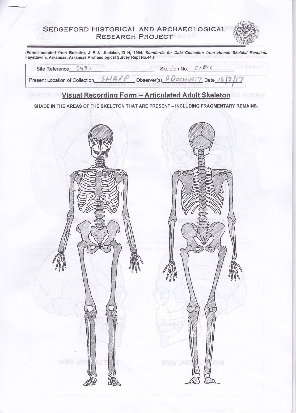
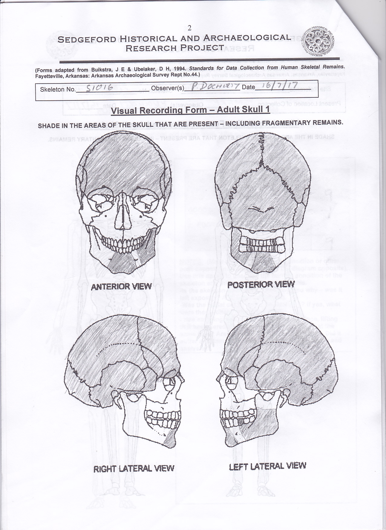
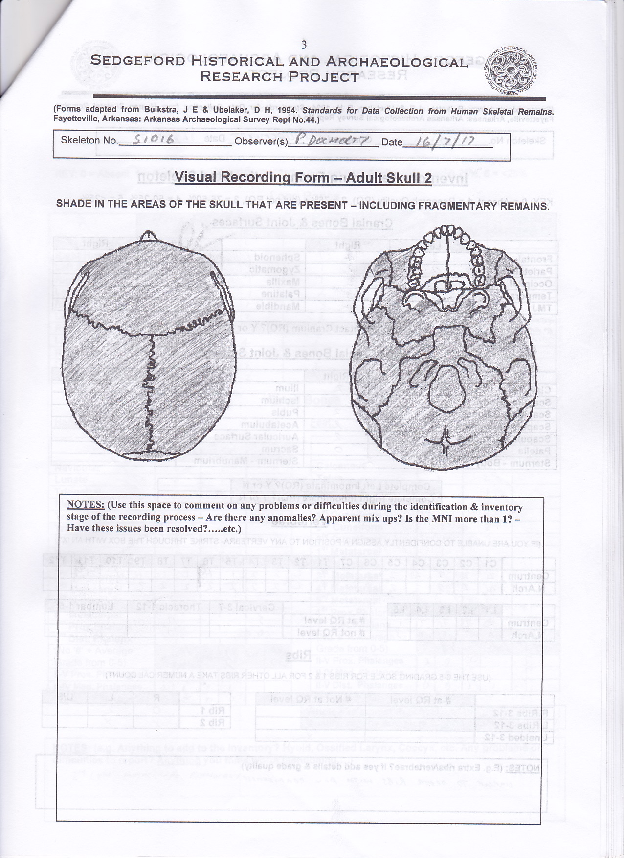
The examination of the skeleton began with the laying out of all bones on a table in a way which represented the anatomical position and orientation relative to the skull. The bones which are present are then shaded these on a series of charts representing range of views within the report.
-
- Skeleton – anterior – posterior
- Skull – anterior – posterior – right and left lateral – superior – inferior
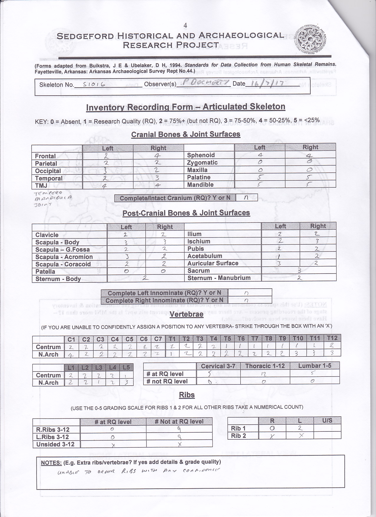
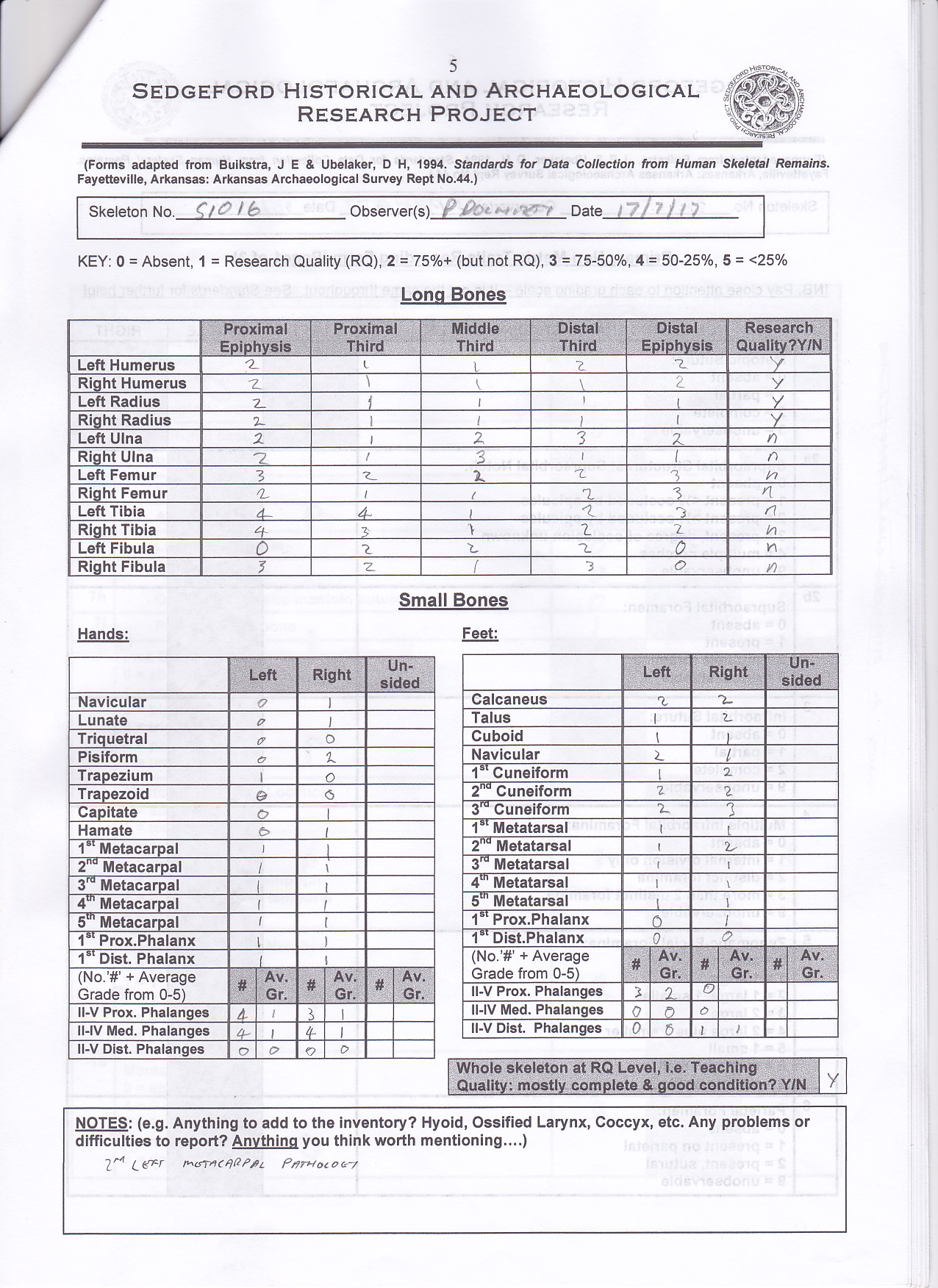
Following on from the initial visual recording a more detailed inventory of all the bones present, including the quality and completeness for each, was recorded using a numerical system as shown in figure 9. The numerical system enables mathematical and statistical queries to be made across the whole database of human remains. The condition of the long bones was recorded in more detail through examination of the proximal, middle, and distal thirds. The quality and presence of the proximal and distal epiphyses was also recorded.
S1016 was in overall good condition with the following observations:
-
- The zygomatic bone along with the maxilla was missing.
- The mandible was fragmented with only two parts extant.
- The cranium had clear evidence of trauma.
- Both patellae were missing.
- The ribs had been broken into several pieces but through identifying the articulating surfaces it was possible to record 9 for each side. The left number 1 rib was also present.
- Both ulnas had been broken.
Primary Non-metric Traits

Non-metric traits, sometimes referred to as discontinuous or discrete traits, are features which are clearly absent or present. With over 400 non-metric traits recognised in the human skeleton they are grouped into seven classes (Mays 1998:102-105):
1. Variations in the number of bones and teeth
2. Anomalies of bone fusion
3. Variation in foramina
4. Articular facet variations
5. Hyperostoses – localised excess of bone formation
6. Hypo-ostoses – local deficiency of bone
7. Variations in the form of the tooth crown
Much of these may be genetic being passed between family and local populations. Some of these may be linked to environmental factors including stress and diet, whilst yet others have been linked to lifestyle i.e. squatting facets. SHARP look at a subset of the full non-metric traits focusing on those in the skull, the septal aperture of the Humerus, squatting facets of the tibia, and the vastus notch on the patella. Regarding S1016 the later features were not present or unobservable as in the case of the missing patellae. The skull had many of the traits unobservable due to the trauma sustained.
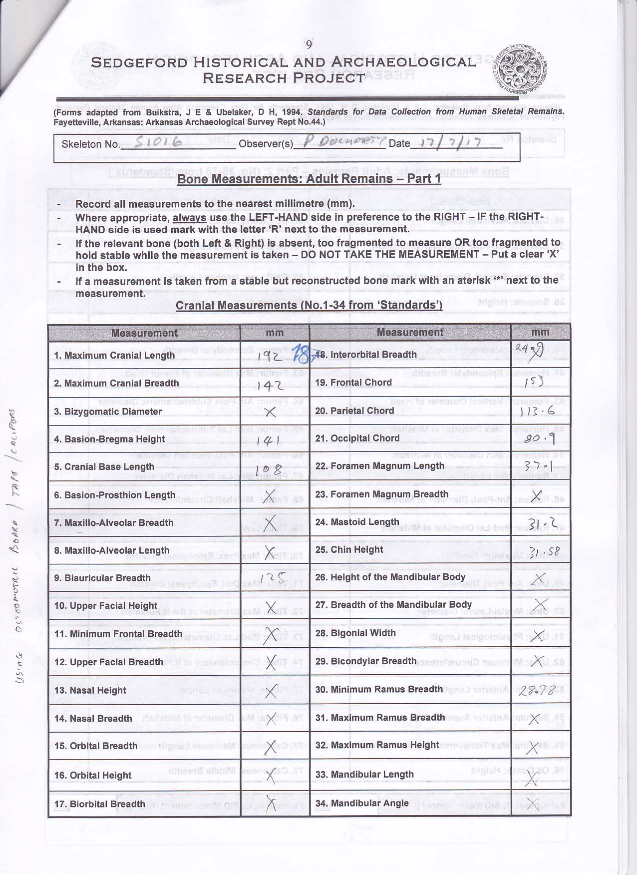
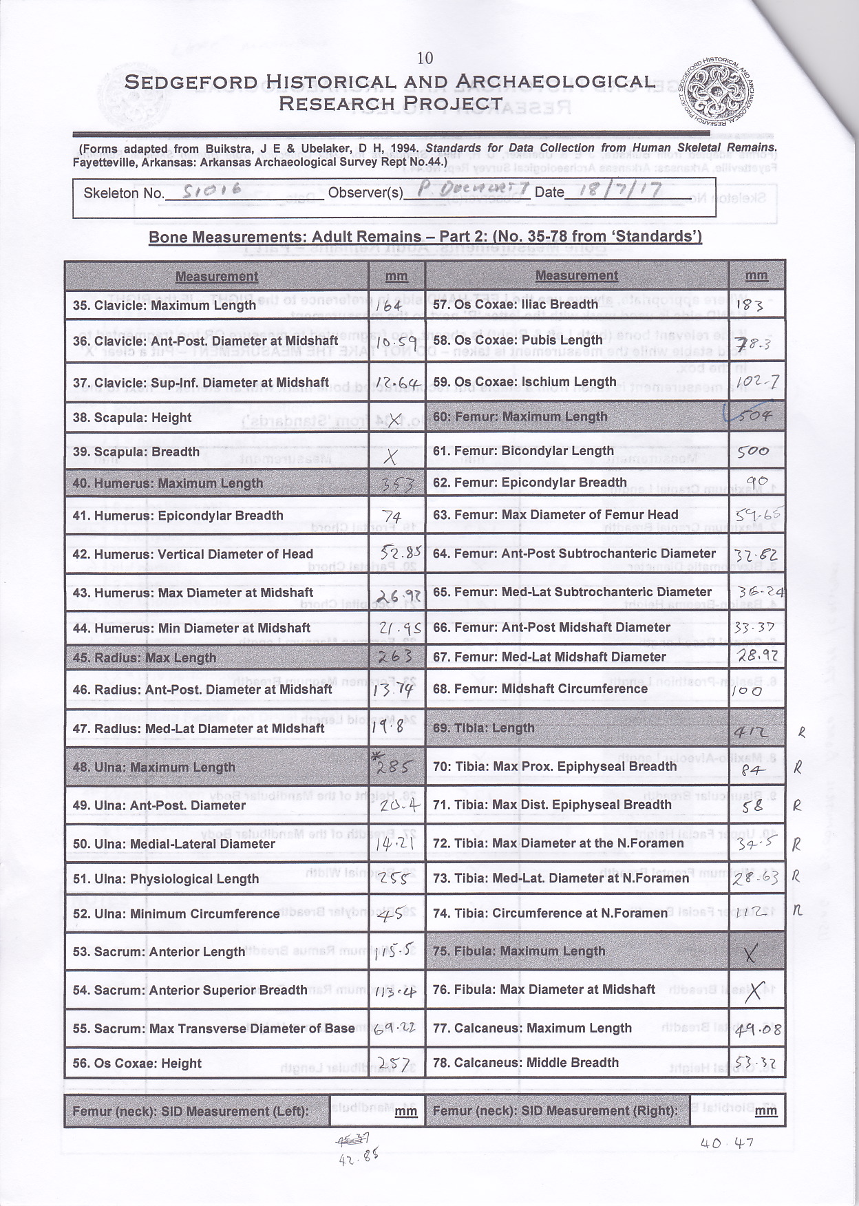
Bone Measurements
All bone measurements were recorded to the nearest millimetre using the left-hand side in preference to the right. If the left-hand side is unavailable, then the right is used but marked with a letter ‘R’ next to the measurement. If neither left or right is present or the remains are too fragmented, then the measurement was not taken and an ‘X’ was recorded. If the bone was reconstructed and stable, then a measurement was taken and an asterisk ‘*’ was recorded alongside the measurement. Measurements were taken from specific skeletal landmarks as identified by Buikstra and Ubelaker (1994). On applying these measurements to S1016, out of the possible 80 measurements, 26 were unobservable and, due to damage present on the left tibia, the right tibia measurements were taken. Whilst both ulnas were broken they could be reconstructed in a stable enough manner to allow for measurements to be taken. The image here shows the left femur length taken using an osteometry board.

Estimation of Stature

Table 1: Stature calculations for skeleton S1016
Stature is estimated by using formula determined by Trotter and Glesser for American white populations (Trotter & Glesser 1952, 1953, 1977). The femur is the most reliable basis for stature estimation followed by tibia, Humerus, and other bones. Table 1 shows the calculations for S1016 giving a maximum height of 187.43cm; the ‘X’ shows that the measurement of that bone was unobtainable. At this stage calculations are presented using both male and female formulae.
Estimation of Age
Calculating the age of the individual can be achieved in several ways. The bones are initially examined for epiphyseal fusion to determine if the individual is an adult; with fusion normally complete by the age of 25-30. In the case of S1016 it was clear that fusion had taken place so age was estimated through examination of the pelvis. Age estimation through the morphology of the pubic symphysis using both the Todd and Suchey-Brooks scales (White & Folkens 2005: 376-379) giving age ranges of 35-39 and 27-66 respectively. In addition, the auricular surface of the ilium was also examined and by using the Lovejoy scale (White & Folkens 2005: 380-383) the age was estimated to be 40-44. Dentition can also be used to estimate age by examining wear on molars. SHARP use Brothwell’s method (Brothwell 1981:72), however for S1016 there was only one molar present for analysis making for a less than ideal estimation resulting in a range of 35-45. By collating the age estimation methods, it was possible that S1016 was aged between 35-44.
Determining Gender

Sex differences in the greater sciatic notch (White & Folkens, 2000, p.417)

Table 2: Bone dimensions as an indicator of sex (Chamberlain, 1994)
Identifying the sex of an individual is established mainly through examination of the pelvis and cranium. Morphology of the ventral arc, sub-pubic concavity, ischio-pubic ramus ridge, greater sciatic notch, and pre-auricular sulcus are used for both the left and right sides where possible. In the case of S1016 these observations indicated strongly towards male.
For the cranium observations of the nuchal crest, mastoid process, supra-orbital margin, glabella, mental eminence, and gonial flaring also indicated a strong male possibility. Bone dimensions can also be used as an indicator of sex see table 2. SHARP also use the supero-inferior femoral neck diameter (SID) and the formula established by Seidemann et al (1998) for Caucasians as follows:
Sex = 0.496 x SID in mm – 15.163
Where sex < 0 is female and sex > 0 is male.
In the case of S1016 the calculation gave a value of 6.0906 for the left SID and 4.91012 for the right SID both strongly indicating male.
Recording Dentition

Dentition Visual Recording Form (SHARP)(A), remaining piece of mandible for S1016 (B-D) (photo: P. Docherty)
Dentition is recorded both visually as a chart (photo part A) and as data giving an indication of the tooth presence, wear, decay (caries), any abscess, and formation of calculus. S1016 had only one tooth present (photo parts C & D), the left lower first molar (19M1). The entire maxilla was missing and the remains of the mandible showed that except for one socket showing premortem loss (17M3) the remaining observable sockets (photo part B) showed post-mortem tooth loss (18M2, 25I1, 26I2, and 27C).
Pathology
Recording the pathology was a descriptive process with care not to analyse too much leading to false assumptions. Observations were methodical beginning at the cranium and working down towards the feet to ensure all bones were examined and recorded. The pathology for S1016 can be seen in table 3 and in following figures. A visual indication of the position of observed pathology is also recorded on a chart.

Cranial trauma observed on skeleton S1016, blow marks from sharp object (A-C) (photo: P. Docherty)

Dentition Visual Recording Form (SHARP)(A), remaining piece of mandible for S1016 (B-D) (photo: P. Docherty)

Post burial modification
To complete the recording of S1016 the overall condition of the skeleton is assessed and any interventions post burial noted. As the excavation took place in August 1997 observations regarding the excavation could not be determined first hand. However, by consulting the original archives at SHARP it was possible to retrieve the site photograph and the burial plan. From the archives, it was noted that the burial of S1016 was part of a mass grave which held at least one other individual, recorded as S1018, with similar pathology and buried during the same period.
The overall condition of S1016 was very good, however during the post excavation analysis a large wedge of bone had been removed from the mid-section of the right femur. Far more than what would have been needed for chemical analysis and this has had the effect of weakening the bone.

Site photograph taken in 1997 showing multiple burials with S1016 on the outside right (image source: SHARP archive)

Plan of multiple burials drawn in 1997 showing S1016 on the right (source: SHARP archive)
Interpretation of the data and possible cause of death
S1016 was a male who was between 172-187cm tall and of solid build. He was aged between 35-44 years old at the time of death and the evidence of skull trauma suggest he was executed by a severe blow to the head. During life, he had been hard working and given the size and strength observed throughout his skeletal frame had eaten a healthy diet. Whilst it is impossible to say with any certainty how S1016 met his fate, given the available data it is possible to offer a plausible scenario. The trauma to the arms is suggestive of two assailants involved in the death and the sequence of events may have played out as follows:
1. The left and right ulnas were both fractured around the mid-third section suggestive of a crossed arm pose in front of the head defending against a weapon strike. Had the break been in the distal third then this would be suggestive of a fighting strike where the opponent would attempt to disarm.
2. The fractures were not clean cut suggesting blunt force consistent with the handle of an axe. The arms likely prevented the axe head from penetrating into the victim’s skull although the presence of a nick can be seen in the front of the skull.
3. The strike is suggestive of a right-handed swing therefore a second assailant may have pulled and twisted the victims left arm generating the spiral fracture in the already weakened bone. This would have had the effect of clearing the head area for the first assailant to strike again.
4. The second strike clearly penetrated the skull in what would have been the death blow.
5. The victim fell over onto his back with his face upwards.
6. One of the assailants then took a sharpened blade and, positioned at the head of the victim facing towards the feet, hacked at the head of the victim numerous times in a side-to-side slashing motion causing severe disfigurement of the face and the jaw to be sliced into pieces.
A similar fate befell S1018 suggesting that these two men were made an example of. This is the working theory of the archaeologists and osteologists analysing the site. Whether the assailants were Vikings in search of supplies and, having come across Sedgeford, decided to exploit their resources cannot be conclusively established. After the initial analysis of S1016 and S1018, dating techniques established the period of burial to be around 800-820 CE which certainly fits in with the early appearance of Vikings on the eastern coast. Out of all the burials excavated so far there have only been 7 individuals who have exhibited signs of violence (Faulkner et al 2014:161) suggesting that Sedgeford was normally a peaceful area.

Re-enactment of the possible cause of death for S1016 presented at the SHARP site tour 21 July 2017 (photo: SHARP)
Report Images
Osteology Report
Osteology Report_0001
Osteology Report_0002
Osteology Report_0003
Osteology Report_0004
Osteology Report_0005
Osteology Report_0006

Osteology Report_0007
Bibliography
Buikstra J. and Ubelaker D. (eds.) (1994) Standards for Data Collection from Human Skeletal Remains. Fayetteville, Arkansas: Arkansas Archaeological Survey Research Series 44.
Brothwell, D. R. (1981) Digging up Bones (3rd edition). London: British Museum Press.
Chamberlain, A. (1994) Human Remains. London: British Museum Press.
Faulkner, N., Rossin, G. and Robinson, K. (eds.) (2014) Digging Sedgeford: A People’s Archaeology. Norfolk: Poppyland Publishing.
Mays, S. (1998) The Archaeology of Human Bones. London: Routledge
Mays, S. (2004) ‘After the bone report: the long-term fate of skeletal collections’, in Brickley, M., and McKinley, J. I. (eds.) (2004) Guidelines to the standards for recording human remains. Southampton, BABAO, Dept. of Archaeology, University of Southampton.
Molleson, T. and Cox, M. (1993) The Spitalfields Project. Volume 2: The Anthropology. CBA Research Report 86, Council for British Archaeology.
Seidemann, R. M., Stojanowski, C. M. and Doran, G.H. (1998) ‘The Use of the Supero-Inferior Femoral Neck Diameter as a Sex Assessor’, The American Journal of Physical Anthropology, 107, pp.305-313.
Trotter, M. and Glesser, C.G. (1952) ‘Estimation of stature from long bones of American whites and Negroes’, American Journal of Physical Anthropology 10, pp.463-514.
Trotter, M. and Glesser, C.G. (1953) ‘A re-evaluation of estimation based on measurements of stature taken during life and of long bones after death’, American Journal of Physical Anthropology 16, pp.79-123.
Trotter, M. and Glesser, C.G. (1977) Corrigenda to ‘Estimation of stature from long bones of American whites and Negroes’, American Journal of Physical Anthropology 47, pp.355-56.
White, T.D. and Folkens, P.A. (2000) Human Osteology (2nd edition). London: Academic Press.
White, T.D. and Folkens, P.A. (2005) The Human Bone Manual. London: Elsevier Academic Press.